Smooth Muscle Diagram Easy / Labeled Cardiac Muscle | Human anatomy and physiology ... : Diagram of contraction of skeletal muscle.
byAdmin•
0
Smooth Muscle Diagram Easy / Labeled Cardiac Muscle | Human anatomy and physiology ... : Diagram of contraction of skeletal muscle.. In this video i have shown the simplest way of drawing muscle drawing. Muscle anatomy shoulder 12 photos of the muscle anatomy shoulder muscle anatomy of the shoulder and neck, muscle anatomy shoulder upper arm, shoulder muscle anatomy game, shoulder muscle anatomy posterior view, shoulder muscle anatomy workout, human muscles, muscle anatomy of the shoulder and neck, muscle anatomy. Occurs in all smooth muscle cells. Diagram of contraction of skeletal muscle. Smooth muscle anatomy smooth muscle tissue is also known as visceral muscle tissue.
Smooth muscle tissue is found around organs in the digestive, respiratory. Smooth muscle anatomy smooth muscle tissue is also known as visceral muscle tissue. Smooth muscle cells lack the striated banding pattern found in cardiac and skeletal muscle, and they receive neural innervation from the autonomic nervous system. However, if you take a little time to learn a few root words, those latin names can give you valuable insights into things like the muscle's size and shape, its location, and even its function. It is found in the walls of ducts and blood and lymphatic vessels, as well as in the walls of the digestive, respiratory and urogenital tracts.
Structure of a Muscle Cell (Muscle Fibre) from www.ivyroses.com 12 photos of the smooth muscle diagram. And smooth muscle in the skin, visceral organs, and internal passageways is essential for moving all materials through the body. This smooth muscle can be found surrounding the walls of the blood vessels, the bronchioles in the lungs, and the sphincter muscles used in the gi tract.the gi tract, which is tubular by design, also houses longitudinal muscles in addition to the smooth. Diagram of contraction of skeletal muscle. • smooth muscles respond to stretch only briefly, and then adapts to its new length • the new length however, retains its original _____ seconds or minutes after it has been elongated or shortened (e.g. Cardiac muscle in the heart wall; Replicate the below figures with labels in your sketchbook. This allows viscera to change diameter from large to almost zero.
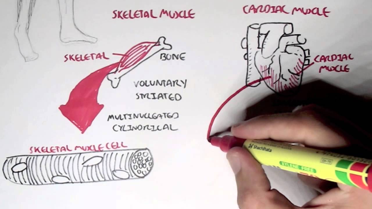
In this video i am gonna to show you how to draw the diagrams of cardiac, straited, smooth muscle for class 1st to 10th.
Wednesday, november 2, 2016 48. The function of smooth muscle tissue; It is layered in a distinctive pattern of circular layers. It is the pen diagram of skeletal, smooth and cardiac muscle for class 10, 11 and 12. Smooth muscle contracts under certain stimuli as atp is freed. Cardiac muscle in the heart wall; Related to the function of movement is the muscular system's second function: Binding of ca2+ to troponin. These cells contain fibers of actin and myosin that run though cells and are supported by frameworks of other many proteins. This allows viscera to change diameter from large to almost zero. It constitutes much of the musculature of (muscle cells are often referred to as muscle fibers because of their narrowness and length.). They range from about 30 to 200 μm (thousands of times shorter than skeletal muscle fibers), and they produce their own connective tissue, endomysium.although they do not have striations and sarcomeres, smooth muscle fibers do have actin and myosin.
They range from about 30 to 200 μm (thousands of times shorter than skeletal muscle fibers), and they produce their own connective tissue, endomysium.although they do not have striations and sarcomeres, smooth muscle fibers do have actin and myosin. It is found in the walls of ducts and blood and lymphatic vessels, as well as in the walls of the digestive, respiratory and urogenital tracts. The maintenance of posture and body position. Muscle anatomy shoulder 12 photos of the muscle anatomy shoulder muscle anatomy of the shoulder and neck, muscle anatomy shoulder upper arm, shoulder muscle anatomy game, shoulder muscle anatomy posterior view, shoulder muscle anatomy workout, human muscles, muscle anatomy of the shoulder and neck, muscle anatomy. On the left is the view with light microscopy.
Popular Jigsaw Games | Page 20 of 34 from proprofs-cdn.s3.amazonaws.com Can be prevented by ca2+ channel blockers. Smooth muscle is defined as a form of muscle tissue that is used by various systems in order to apply pressure to vessels and the organs.the smooth muscles are made up of sheets or strands of smooth muscle cells. A smooth muscle can contract more than 2/3rd its stretched length while skeletal muscle contract up to 1/3rd. Smooth muscle tissue, unlike striated muscle, contracts slowly and automatically. In this video i am gonna to show you how to draw the diagrams of cardiac, straited, smooth muscle for class 1st to 10th. Look at the name of the muscle for clues. The smooth muscle contraction is much slower than in the striated muscle primarily due to the presence of g protein coupled ligand receptors instead of ion channel coupled ligand gated receptors present in striated muscle. This type of smooth muscle is observed in the large airways to the lungs, in the large arteries, the arrector pili muscles associated with hair follicles, and the internal eye muscles which regulate light entry and lens shape.
And smooth muscle in the skin, visceral organs, and internal passageways is essential for moving all materials through the body.
Marked shortening of smooth muscle durin contraction. Muscles are the only tissue in the body that has the ability to contract and therefore move the other parts of the body. Replicate the below figures with labels in your sketchbook. Smooth muscle is widely distributed in the body. Smooth muscle in the walls of arteries is a critical component that regulates blood pressure necessary to push blood through the circulatory system; These cells contain fibers of actin and myosin that run though cells and are supported by frameworks of other many proteins. Mesenchymal stromal cells are required for regeneration and. Smooth muscle, muscle that shows no cross stripes under microscopic magnification. 3 types of muscle tissue. These cells have fibers of actin and myosin which run through the cell and are supported by a framework of other proteins. It is the pen diagram of skeletal, smooth and cardiac muscle for class 10, 11 and 12. Smooth muscle is a type of muscle tissue which is used by various systems to apply pressure to vessels and organs. This type of smooth muscle is observed in the large airways to the lungs, in the large arteries, the arrector pili muscles associated with hair follicles, and the internal eye muscles which regulate light entry and lens shape.
Replicate the below figures with labels in your sketchbook. And smooth muscle in the skin, visceral organs, and internal passageways is essential for moving all materials through the body. Marked shortening of smooth muscle durin contraction. The pupillary sphincter muscle in your eye is a smooth muscle that shrinks the size of your. Smooth muscle tissue is found around organs in the digestive, respiratory.
Myology - Introduction (Skeletal, Cardiac, Smooth Muscles ... from i.ytimg.com Smooth muscle is composed of sheets or strands of smooth muscle cells. A smooth muscle can contract more than 2/3rd its stretched length while skeletal muscle contract up to 1/3rd. In this video i have shown the simplest way of drawing muscle drawing. It is the pen diagram of skeletal, smooth and cardiac muscle for class 10, 11 and 12. Replicate the below figures with labels in your sketchbook. Binding of ca2+ to troponin. Simple smooth muscle diagram labeled written by jupiterz tuesday, december 12, 2017 1 comment edit. These cells have fibers of actin and myosin which run through the cell and are supported by a framework of other proteins.
Can be prevented by ca2+ channel blockers.
Smooth muscles have a much stronger ability to contract than skeletal muscles, and are able to maintain contraction longer. Can be prevented by ca2+ channel blockers. Like cardiac muscle, smooth muscle is involuntarily controlled. Mesenchymal stromal cells are required for regeneration and. Smooth muscle in the walls of arteries is a critical component that regulates blood pressure necessary to push blood through the circulatory system; Replicate the below figures with labels in your sketchbook. Smooth muscle is composed of sheets or strands of smooth muscle cells. It is layered in a distinctive pattern of circular layers. When you finish with the above photo, head to the following model on posemanics and draw and label the linked model. Smooth muscle is defined as a form of muscle tissue that is used by various systems in order to apply pressure to vessels and the organs.the smooth muscles are made up of sheets or strands of smooth muscle cells. Muscles are the only tissue in the body that has the ability to contract and therefore move the other parts of the body. Smooth muscles in a woman's uterus (or womb) help to push babies out of the body during childbirth. Smooth muscles exhibits a phenomenon called _____ in which:
The pupillary sphincter muscle in your eye is a smooth muscle that shrinks the size of your smooth muscle diagram. Smooth muscle in the walls of arteries is a critical component that regulates blood pressure necessary to push blood through the circulatory system;